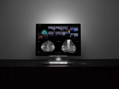
What is a breast imaging display?
A breast imaging display is a medical-grade monitor that meets the high demands of breast imaging. It is used by breast radiologists to view breast images such as mammograms and breast tomosynthesis slices. Breast imaging displays come with special tools and technologies to help radiologists improve the detection of breast cancers.
Acquisition vs display
According to the ACR (American College of Radiology), the resolution of a breast imaging display should match the resolution of the imaging (or acquisition) system as closely as possible.*
Only then, enough of the picture elements – meaning important image details such as subtle masses and calcifications – will be clearly visible on the screen. Mammography monitors with a resolution between 5 megapixel and 12 megapixel best support the ACR guidelines.
* ACR–AAPM–SIIM PRACTICE PARAMETER FOR DETERMINANTS OF IMAGE QUALITY IN DIGITAL MAMMOGRAPHY, revised 2017
Mammography monitors: more screen, fewer clicks
Breast imaging displays with additional vertical resolution help breast radiologists fit more of the breast image on the display. Since the specified practice guideline in breast imaging is that all images should be viewed at 1:1 or 100% size*, this is important for a radiologist’s reading workflow. Additional resolution will save radiologists a number of clicks per day when getting the best image for analysis, by minimizing panning and zooming and reducing windowing and leveling time.
*ACR–AAPM–SIIM PRACTICE PARAMETER FOR DETERMINANTS OF IMAGE QUALITY IN DIGITAL MAMMOGRAPHY, revised 2017

High-resolution mammography monitors with more vertical resolution optimize breast imaging workflow by reducing the number of steps needed for viewing breast images full size (1:1 reading). As you can see in the image above, a standard 5MP display requires 4 steps (clicks) to read the image, a 5.8MP display requires only 2 steps.
Fusion increases productivity
Breast imaging displays can be Fusion displays, which combine real estate of two diagnostic displays into one. Breast radiologists can lay out multiple images anywhere on the screen, for easy side-by-side comparisons.

A mammography display that supports flexible color multimodality imaging. 2D, breast tomosynthesis, ultrasound and breast MR images can be laid out anywhere on the screen.
2D vs 3D mammography
3D mammography, also known as digital breast tomosynthesis, takes multiple images (or slices) from the breast to create a 3D image. It can take more time to diagnose digital breast tomosynthesis studies because radiologists must scroll or cine through many images. At the same time, it is more challenging to clearly detect microcalcifications in moving images. Special mammography displays can counter these challenges, ensuring crisp and in-focus moving images with no blurring.

Higher brightness, better detection
Digital breast images require the highest resolution and brightest displays for review. Because the higher the brightness, the bigger the chance to catch breast cancer. Studies have shown that calibrated luminance levels of 1000 cd/m² lead to increased detection probability of breast microcalcifications, compared to calibrated luminance levels of 500 cd/m².
*Kimpe, T. R. & Xthona, A. (2012). Quantification of Detection Probability of Microcalcifications at Increased Display Luminance Levels. Breast Imaging, Springer 7361, 490-497. 2012.
Mammography monitors and reading ergonomics
Tasked with reading dozens of studies per day in an environment with low levels of ambient light, breast radiologists often experience eye fatigue. Breast imaging displays offer a high brightness and can control ambient light to ensure optimal reading conditions.
Another major cause of discomfort is neck strain, which results from frequent head movements when viewing images on multiple screens. Some breast imaging displays have been specifically designed to present images within the optimal field of vision to maintain effortless visual acuity.

A diagnostic display is designed to mirror a human's natural field of vision to minimize head, neck and eye movement.
Conclusion
In digital breast imaging, whether 2D mammography or digital breast tomosynthesis, the quality of the medical display has a direct impact on the decisions breast radiologists make. Providing breast radiologists with a breast imaging display that helps them improve breast cancer detection, speed up workflow and read more comfortably is a vital factor in the correct interpretation and diagnosis of breast cancer.

